Technology
Why We Scan in 3D Before Every Implant
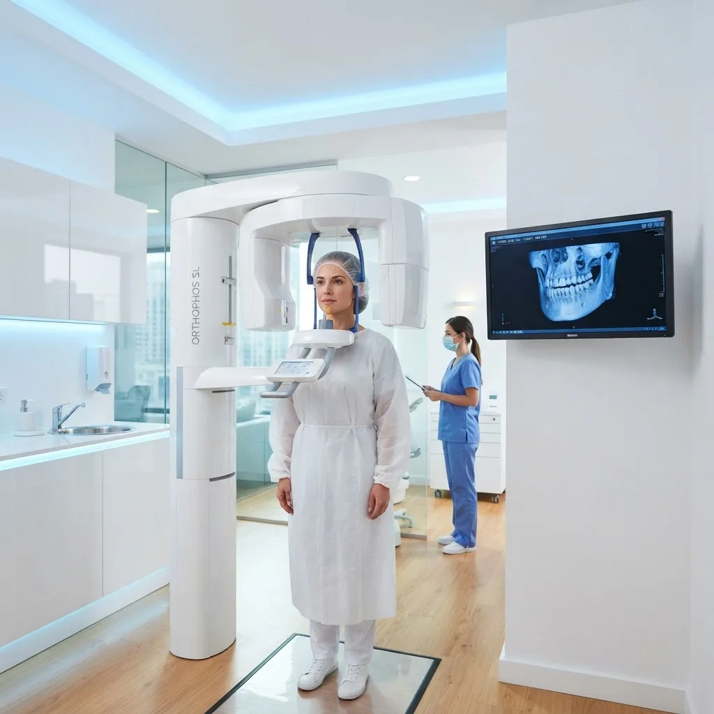
A traditional dental X-ray is a flat picture of a space that is not flat. It tells us a tooth is there and roughly how deep the root goes, but it flattens everything in front of and behind that tooth into a single grey image. For a filling, that is enough. For an implant, it is not.
What a cone beam scan actually shows
A cone beam scan takes roughly twenty seconds and produces a volume rather than a picture. We can rotate it, slice it at any angle, and measure real distances in millimetres. Three things matter most before an implant:
- Bone height and width at the exact site, which decides whether an implant fits or whether a graft comes first.
- Bone density, which determines how much healing time you need before the crown goes on.
- The position of the inferior alveolar nerve in the lower jaw, which is the structure we most need to avoid.
Why it changes the surgery
Once we have the volume, we plan the implant on screen before you are in the chair. We choose the length, width, and angle, then check the position against the nerve and the neighbouring roots. If the plan does not work, we find out at a desk instead of mid-procedure.
That plan can then be printed as a surgical guide that sits over your teeth, so the drill enters at the angle we decided in advance. The appointment gets shorter, the site stays smaller, and recovery is usually easier because there is less exploring involved.
What about the radiation?
It is a fair question and one we get often. A dental cone beam scan is a fraction of the dose of a medical CT, and we only scan the region we are treating rather than the whole head. We still only take one when the result will change what we do, which for implants it reliably does.